Posted on November 29, 2003 by Inception Fertility
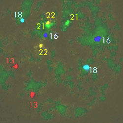
PGD FISH (Fluorescent in situ hybridization) Photo:An embryo with normal chromosomesWe are looking here at the DNA from a cell taken from a human embryo. It has been stained green, and colored fluorescent probes have been applied, which are specific to individual chromosomes. This allows us to count the number of chromosomes and tell if the embryo is normal or not for those chromosomes tested. For the chromosomes that we are interested in counting, we should see 2 brightly colored spots, since we have 2 copies of each chromosome. In this picture, we see 2 red spots (= 2 copies of chromosome 13), 2 yellow (chromosome 22), 2 light blue (chromosome 18), 2 dark blue (chromosome 16) and 2 green (chromosome 21). This embryo has the correct number of these chromosomes. Note that one of the green spots and one of the yellows are very close together in the picture, but they are definitely there.
Welcome to the Pacific Fertility Center Blog! Nationally and internationally recognized for providing exceptional reproductive care, our team believes in empowering people with the knowledge they need to navigate their unique fertility journeys.
From information on the latest fertility treatments to valuable insights on egg donation, surrogacy, and everything in between, the Pacific Fertility Center Blog is your ultimate resource for all things reproductive care and support. Read on to learn more, and contact us today if you have any questions or want to schedule a new patient appointment.