Fibroids
Fibroids of the uterus (leiomyomas or myomas) are benign (non-cancerous) tumors made of smooth muscle that usually grow within the uterine walls. Fibroids may cause heavy menstrual flow, severe cramping, pelvic pressure and bladder or bowel problems. Depending on their location, size and growth rate, fibroids can also interfere with conception and pregnancy.
Diagnosing Fibroids
A variety of means are available for diagnosing fibroids, depending on their size and location.
- Pelvic Examination. If the fibroid tumor or tumors are very large, they may be felt on pelvic examination.
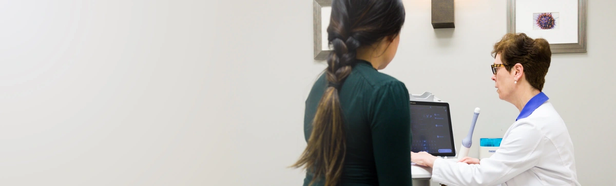
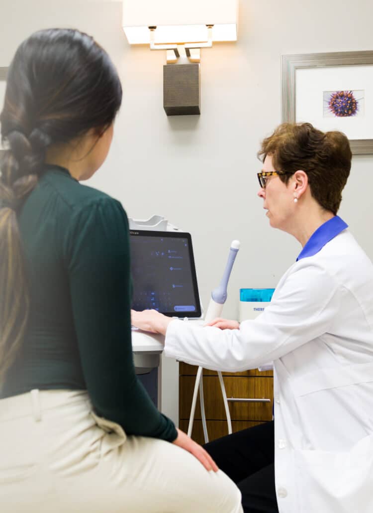
- Routine Pelvic Ultrasound. This is a transvaginal ultrasound, used to view the lining of the uterus. It is usually the best way to see a fibroid. For the ultrasound, the doctor inserts a slim probe called a transducer into the vagina. In order to evaluate the potential effect on the uterine cavity, the ultrasound should be performed shortly before ovulation, when the endometrial lining is at its thickest.
- Hysterosonogram Ultrasound (Saline sonogram or water sonogram). This is a vaginal ultrasound in which the physician injects sterile saline (salt water) into the uterine cavity. The saline serves to enlarge the uterus, and also makes it easier to view and measure any fibroids within the area. It is best used to visualize submucous fibroids, those within the uterine cavity.
- Hysteroscopy. Hysteroscopy is a minor surgical procedure that involves placing a lighted telescopic instrument (hysteroscope) through the uterine cervix and visualizing any abnormalities within the wall of the uterine cavity. If a fibroid is seen, it can sometimes be removed during the same procedure.
- Magnetic resonance imaging MRI. This procedure uses a powerful magnetic field, radio frequency pulses and a computer to produce highly detailed pictures of the soft tissue (as well as virtually all other body structures). In the case of fibroids, MRI can help to delineate multiple growths of various sizes in order to insure that all may be removed. Because of its specificity, the MRI can help to distinguish fibroids from the one other uterine abnormality that it can sometimes be confused with, adenomyosis. Adenomyosis cannot be removed surgically, however fibroids can – thus the importance of distinguishing between the two.
Treating Fibroids
In a surgical procedure called myomectomy, the fibroid can be removed, preserving the uterus and ovaries so that conception can occur. There are various methods to perform myomectomy, depending on the location, size and number of fibroid(s).
- Myomectomy with laparotomy. If the fibroids are mostly within the wall of the uterus, the surgery is usually done by laparotomy, involving a large incision through the abdominal wall. This "abdominal myomectomy" is the best approach for large and/or multiple fibroids within the muscle wall of the uterus. This surgical procedure can be performed by most all gynecologists. Of course, as this is a major abdominal surgery, a hospital stay of 2-3 days is usually required and recovery can take 4-6 weeks. Most women who have an abdominal myomectomy will need to have a Cesarean section with any subsequent pregnancy.
- Myomectomy with a hysteroscope (hysteroscopic resection). If the fibroid is mostly situated within the uterine cavity, the best approach is usually through a hysteroscope, a thin lighted telescope inserted through the vagina and into the uterus. The hysteroscope is fitted with specific surgical instruments, enabling the physician to view and remove the fibroids. This surgery must be done by gynecologists with adequate training in hysteroscopy. As no incisions are involved it is usually performed as a day surgery with no overnight hospital stays and recovery is rapid – less than a week - and there are no incisions.
- Myomectomy with a Laparoscope (Laparoscopic myomectomy). This procedure uses a laparoscope and one or more small incisions in the abdomen. Though laparoscopic myomectomy is minimally invasive, it requires special training by a gynecologic surgeon. In recent years, laparoscopic surgeons are also starting to use robotic assisted surgery and this has been especially helpful for myomectomies. There are only a handful of gynecologists adequately trained in robotic laparoscopic myomectomy.
With laparoscopic myomectomy, scar tissue formation after surgery is usually less than with open surgery; and recovery time is relatively quick, due to minimal abdominal incisions. - Uterine Artery Embolization. This relatively new procedure holds promise, but because long term effects are still being studied, it is not yet recommended for women who want to preserve their fertility. The embolization procedure blocks fibroid blood supply by injecting small particles (beads) into the arteries that supply the fibroids. For the embolization, the physician guides a catheter through the leg arteries and into the uterine artery.
Endometrial Polyps
Small, benign overgrowths of the normal tissue lining the uterus, the endometrium. Many women have polyps but may have no symptoms and never know they have them unless they happen to have a pelvic ultrasound. They are very common in women in their 30's and 40's and can probably cause infertility.
- Diagnosing Polyps. Routine vaginal ultrasound performed just prior to ovulation is usually sufficient to diagnose a polyp. If unclear, a hysterosonogram or hysteroscopy (see above under Diagnosing Fibroids) can be performed. Endometrial polyps are often missed on hysterosalpingograms because the radio-opaque dye blocks their visualization.
- Treating polyps. Endometrial polyps are successfully treated with hysteroscopy (see above) and the patient can try to conceive immediately after the polyp is removed.
Uterine Scar Tissue
Sometimes, particularly after a dilation and curettage procedure, a woman may develop scarring of the uterine cavity. If severe enough scarring is present, this is known as Asherman's syndrome. This type of scarring may make it difficult or impossible for an embryo to implant in the uterus.
-
Diagnosing Uterine scarring. Ultrasound, hysterosonogram, hysteroscopy or hysterosalpingography can be used to diagnose intrauterine scar tissue.
-
Treating Uterine scarring. Hysteroscopy with resection of the scars.
Uterine Congenital Anomalies
In rare cases, women may be born with a uterus that has a vertical septum (septate uterus) or even a duplication of the uterus (uterine didelphys). Depending on the severity, surgery may be recommended.
- Diagnosing Uterine Congenital anomalies. Ultrasound, hysterosonogram, hysteroscopy or hysterosalpingography can be used to diagnose any of these types of uterine anatomic disorders.
- Treating Uterine anomalies. Hysteroscopy with resection of the septum is the treatment of choice for a septate uterus. For a uterine duplication, surgery is not usually required but avoidance of twins is very important because of the much greater risk of pre-term labor and pre-term delivery.
At our Northern California San Francisco Bay Area clinic, PFC's fertility doctors offer a wide variety of treatments to effectively treat fibroids; including those listed above.
Would you like to learn more about fibroids and fertility? Join us for our next free fertility seminar!
Each month, Pacific Fertility Center® hosts a complimentary educational seminar hosted by our nationally renowned 'Top Docs'. Seminar attendees have the opportunity to tour our San Francisco Bay Area fertility clinic, and ask our doctors fertility questions.
Some common fibroid questions include:
- Which fibroid treatment will best improve my chances of getting pregnant?
- Will I need to have a C-section if I get pregnant after fibroid removal surgery?
- I have many choices in Northern California's San Francisco Bay Area. Why should I choose PFC?
- And much more... We're here to answer your questions.
Pacific Fertility Center is located right next to Fisherman's Wharf and the Embarcadero in the San Francisco Bay Area, with convenient access from many Northern California communities. Sign Up for the next new patient seminar @PFC »